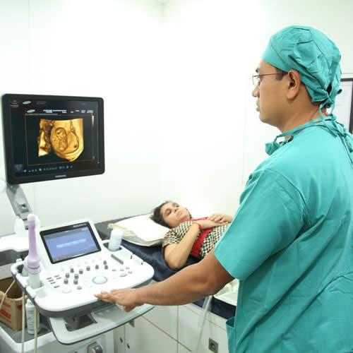
Ultrasonography
Medical ultrasound (also known as diagnostic sonography or ultrasonography) is a diagnostic imaging technique based on the application of ultrasound. It is used to see internal body structures such as tendons, muscles, joints, vessels and internal organs. It’s aim is often to find a source of a disease or to exclude any pathology. The practice of examining pregnant women using ultrasound is called obstetric ultrasound, and is widely used.
Ultrasound is sound waves with frequencies which are higher than those audible to humans. Ultrasonic images also known as sonograms are made by sending pulses of ultrasound into tissue using a probe. The sound echoes off the tissue; with different tissues reflecting varying degrees of sound. These echoes are recorded and displayed as an image to the operator.
Role of Ultrasound In IVF Treatment
Ultrasound is the most versatile method for pre-treatment assessment in IVF being the dominant instrument for assessing ovarian reserve, pelvic pathologies and for assessing the uterine cavity. The ability of ultrasonography to measure endometrial thickness in addition to detecting uterine masses gives it an edge over laparoscopy/hysteroscopy as a diagnostic procedure in uterine cavity assessment, although hysteroscopy has the advantage of therapeutic potential. Similarly, ultrasonography is superior to biochemical methods for follicular monitoring because of its ability to demonstrate the number and sizes of follicles, and guide preparations for oocyte retrieval. The relative ease of ultrasound guided oocyte retrieval; its less technical demands and the possibility of conducting the procedure under local anaesthesia have made ultrasound guided oocyte retrieval more popular across the world. Randomized controlled trials show that ultrasound-guided transfer techniques have better outcomes than the clinical touch technique in terms of on-going pregnancies and Clinical pregnancies. Ultrasonography is now the key instrument for diagnosing and monitoring pregnancy following embryo transfer, biochemical methods being complimentary.
3D ultrasound for predicting endometrial receptivity in ARTs
The term “uterine receptivity” refers to a state when endometrium allows a blastocyst to attach, penetrate and induce changes in the stroma, which results in the so-called process of implantation.
With the advent of three-dimensional ultrasound it became possible to perform a reliable and reproducible sonographic endometrial volume calculations as well as an assessment of endometrial and subendometrial vascularization. Therefore, some researchers have evaluated the role of endometrial volume as well as subendometrial and endometrial vascularization for predicting uterine receptivity.
What if I need frequent scans through my pregnancy?
You may be offered more scans than usual to monitor the growth and well being of your baby if you:
- had complications in a previous pregnancy
- have diabetes
- have high blood pressure
- have a BMI of over 30
- are expecting twins or more
- have any complications in your present pregnancy
But even if you have more frequent scans than other expecting mums, there is no greater risk to you from the scans than to others. On the contrary, if your doctor is prescribing extra scans, it is because the information she collects from these scans helps her to ensure that you and your baby are doing well. The benefits from serial scans in monitoring pregnancies are considered to outweigh any potential risks.